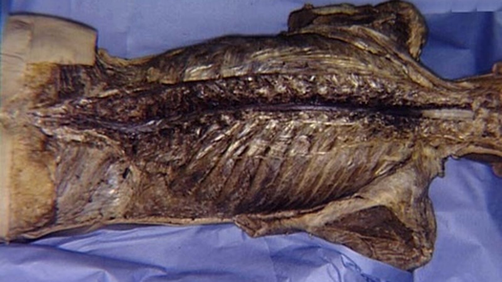
James Clark is back to walking after severing his spinal column. James Clark to walk again after a car accident left him with a severed spine.
In 2012, Illis[] remarked: “There is some evidence of CNS regeneration, but the evidence is of doubtful neurological significance. There is not a single example of experimental work translating into a therapeutic effect…It would be difficult to find any other branch of science with over a century of such sterile endeavour. In effect, there has been repetition of the same idea, albeit with different techniques, that is, looking at the lesion site. Are we sentenced to repeating the same experiments in the hope of expecting a different result?”
Is Illis right?
Clinical trials of a wide variety of different cell lines implanted at or around the lesional level (Schwann cells, olfactory ensheathing cells, mesenchymal/stromal stem cells, multipotent progenitor cells, neural stem/progenitor cells, embryonic stem cells, umbilical cord blood cells) have been conducted (and many others are in progress: see at ClinicalTrials.gov) – no biological cure defined as independent, unaided deambulation has been achieved to date. Some open-label, uncontrolled reports claimed positive effects, even years after the injury, with some patients walking again with braces and support (although not restitutio ad integrum).[,] However, negative studies and complications are equally on record.[,] Scaffolds in combination with cell grafts for chronic spinal cord injury (SCI) have been implanted, but early results do not seem especially promising.[] In sum, while some benefit may accrue from cell grafts, they alone cannot cure paralysis.[]
More than 50 years ago Walter Freeman suggested the severance-reapposition model for chronic SCI; he removed the damaged cord in dogs creating a gap, performed a complete en bloc vertebrectomy[] thus shortening the spine, brought the two fresh cord stumps in contact with fresh plasma and sutured the dura tightly: walking animals resulted after several months.[,10,11,] He also experienced recovery of gait in mice and rats submitted to full transection. Spinal cord transection is rare in the clinic, but has been around as a model of SCI[] for more than 150 years, i.e. since the first report of motor recovery in pigeons by Brown-Sequard in 1848.[33] In 1894, Stroebe[34] described the presence of regenerating nerve fibers in the scar tissue between the ends of severed spinal cord in rabbits (similar to Freeman). Dogs showed recovery of locomotive ability in the hindlimbs to stand and walk after complete transection of the spinal cord at the lumbothoracic level in some studies.[7,13,15] However, much of this recovery has been ascribed to reflex motor activity, as suggested by Sherrington. For instance, Handa et al.[] performed a complete transection of the midthoracic (T9 or T10) spinal cord on 9 adult female dogs. Follow-up lasted 6–39 months. Within several weeks, muscle tone of the hindlimbs was gradually increased accompanied by development of flexion reflex with after-discharge in addition to monosynaptic reflexes. Alternating stepping movement also began to develop. Afterward, extensor thrust and crossed extension reflex were observed. Standing behavior of the hindlimbs was found after sufficient development of the extensor thrust and correct placement of the pads of the toes. Steady development of stepping and standing caused forward locomotion using fore- and hindlimbs; 7 out of 9 could walk on open ground. This ability of locomotion by the hindlimbs of the spinal dogs reached a plateau 6 months after the surgery. Walking behavior of the hindlimbs was not inhibited by additional spinal cord transection in the 2 dogs where it was done, pointing to spinal automatisms and development of responses induced by afferent inflow from outside the cord as the reason for such functional recovery. This was also corroborated by electrophysiological absence of conduction across the transection. However, Freeman observed direct electrophysiological conductance across the apposed stumps.[,] Complete dorsal transection in mice has been followed up in one natural history study: at 7 days, 33% of them displayed weak nonbilaterally alternating movements (NBA). At 14 days, increased NBA were observed and the first bilaterally alternating movements (BA) in 10% of the mice. A progressive increase of movement frequency and amplitude was found after 2–3 weeks. By the end of the month, 86% displayed mixed NBA and BA. However, none of them recovered the ability to stand or bear their own weight with the hindlimbs.[] Freeman observed recovery after a much longer follow-up.[,]
In any case, what is clear is the extremely long time required for recovery to materialize (translated to the clinic, years). In fact, case reports of patients in whom the injured segment has been removed and treated locally took at least 1.5 years (up to 3) to reacquire partial aided locomotion.[] This lag must be dramatically shortened to justify clinical trials.
Spinal shortening and stump reapposition requires an exact understanding of what takes place at the time of the section. Yoshida et al.[] studied this model in the rat. The sharpness of the transection turned out to be one of the most important factors for successful axonal regeneration. An extremely sharp transection produced edema-free lesions and later formed neither cysts nor scars, whereas a relatively blunt transection produced edema followed by scars and cysts around the lesions. Consequently, the spinal cord was transected using the edge of a razor which was as sharp as possible to minimize traumatic injury. However, the stump of the spinal cord resulted in edema since it took 10 or 20 min to bring together the stumps of the spinal cord following transection. This dovetails with a rodent study: the ends of the transected spinal axons remain stable for only about 10–20 minutes before they undergo fragmentation (the first step before classic Wallerian degeneration, or dieback) at both ends spanning 0.3 mm, only to stabilize and persist for 3–7 days; however, about 30% of proximal axons then start growing again within 6–24 hours.[] Older studies showed that immediately following transection of the spinal cord axoplasm escapes from both the proximal and distal portions of some of the cut axons: the extent of axoplasmic loss is generally greater in larger myelinated fibers. In contrast, the small fibers, whether myelinated or unmyelinated, show little if any loss of axoplasm. One hour after the transection, the proximal and distal ends of the axons have retracted from the transection site, and both ends are separated by 1–2 mm or more from the transection site. The axoplasmic leakage stops within a few hours of the transection. Electron microscopic observations indicate that the tip of an axon is lined by axolemma within 1 hour; in addition, layers of collapsed myelin form a septum in front of the axonal tip. At about 3 hours after axonal transection, the axon becomes swollen and irregular in shape and massive accumulation of lysosomes and release of autolytic lysosomal hydrolases is observed within both the rostral and the caudal spinal cord stumps, peaking at 3–7 days and declining at 14 days: cavitation is the result.[,22,] Both the proximal and distal ends swell because axoplasmic transport is bi-directional. Degeneration spreads in both directions along the axon from the transection site, but only for a short distance in the proximal portion: in a clean cut, only one or two internodes may be involved within the proximal stump.[3] In the distal axon, however, Wallerian degeneration occurs. In view of this data, it is obvious that whatever treatment must be brought to bear immediately or within minutes (less than 10), and we suggested sectioning the cord beyond the point of actual fusion and further trimming the stumps at the last moment before reapposition.[]
Having defined a temporal relationship between section, apposition and deployment of therapy, one has to select the best alternative. Almost all animal studies of cord transection (including the current SNI study),[] deployed the studied experimental procedure immediately after section and are thus relevant to this discussion.
Faraway 3 arctic escape gameplay pc. After you put plate in the square space and activate portal, you’ll see the symbol on the plate pulsing, like it’s got power now. Don’t enter portal yet.
Cell grafts of the kind discussed above have been assessed in a large number of animal studies. To establish the most effective graft, one has to compare the reported outcomes of full transection studies and for the past 20 years, the 21-point Basso–Beattie–Bresnahan (BBB) scale has been employed in most rodent studies for this purpose (0 = paralysis of hind limbs, 21 = normal gait). Scores from 1 to 7 (LEVEL 1) mark the return of isolated movements of hip, knee, and ankle, scores from 8 to 13 (LEVEL 2) the return of hindlimb coordination, and scores from 14 to 21 (LEVEL 3) the recovery of predominant paw position, trunk stability, and tail position. As mentioned above, BBB scores of up to 2 (rarely up to 5) can be seen in untreated rats. However, while scores up to 5 may not be considered recovery, actually several reportedly positive studies did not go beyond 5. With this frame in mind, a literature review reveals that the vast majority of studies did not go beyond LEVEL 2, with many not passing LEVEL 1.[,] The latest study published in this journal with xenogenic mesenchymal stem cells (xenoMSC) is in this range.[] The only outlier is the study reported by Ziemlinska et al.,[] exploiting BDNF overexpression via gene vectors injected bilaterally at the level of the transection. Assessed on a modified BBB scale (22 points), treated rats reached a score of 13.7 (up to 18), i.e., LEVEL 3 after a few weeks! In conclusion, cell grafts alone are not the key to fast reconstruction of the transected cord (BDNG gene therapy remains to be confirmed).
These results must be compared with the local application of the much less expensive and widely available fusogens such as PEG. In the latest rodent study, on day 28, the mean BBB score of the PEG group was 12 (range: 7–20, median 12) vs 4.4 (range: 3–5, median 5) in controls. Two rats reached 19 and 20,[] which is better than all published cell grafting studies to date. Parenthetically, in the SNI study, two rats reached a maximum score of 12.
Where does this leave us?
To treat an injured cord, the injured segment must be removed. Two options are possible: shorten the spine and the cord (via a vertebrectomy or multiple diskectomies) (even a 1 cm slice should suffice in some cases, as shown by Freeman),[,] and fuse the two stumps or fill the gap. This latter has been done but it takes well over 1 year or more to see partial effects,[] given the long gap regrowing fibers must cover. Thus, a clinical trial of PEG-assisted spinal shortening is warranted. Of course, auxiliary measures to improve results are also indicated, ranging from electrical stimulation of the fused segment to electroacupuncture (as seen in rodent transactions), intermittent ischemia, and others. Neuroengineered electrical bridges are in the works, but still some way off.[,]

